Blood Vessels Labeled Brain / Cerebral Circulation Adaptations Teachmephysiology : Veins return blood back toward the heart.. Supplies the posterior brain, blood supply to the entire brain is ensured by anastomoses between the vessels. In the cerebral medulla, the arteries and veins of the right side of the body are controlled from the left side of the brain; Blood in the brain is supplied by two pairs of large blood vessels (arteries): The dense tight junctions between endothelial cells prevent paracellular transport through the. The capillaries also connect the branches of arteries and to.
Supplies the posterior brain, blood supply to the entire brain is ensured by anastomoses between the vessels. The brain and its surrounding blood vessels exist in a close relationship. The two cell types ensure the integrity of the neural vasculature by maintaining the blood. A blood clot that lodges in a brain blood vessel, causing a stroke. Blood flows throughout the body tissues in blood vessels, via bulk flow (i.e., all constituents together and in one direction).
Cerebral Artery Images Stock Photos Vectors Shutterstock from image.shutterstock.com The dense tight junctions between endothelial cells prevent paracellular transport through the. Veins return blood back toward the heart. This is particularly important structure due to its clinical implications, which are discussed in more detail in the article. Internal carotid artery (anterior circulation), vertebral artery (posterior circulation), and their hexagonal anastomotic network called blood brain barrier refers to the wall between the brain tissue and blood vessels. • identification of blood vessels as arteries, capillaries or veins from the structure of their walls. Fill in the blanks with the appropriate words to describe blood flow from the heart. Endothelial cells are labeled in red and pericytes in green. Equal to the intestinal muscles that move the food morsel along brain level:
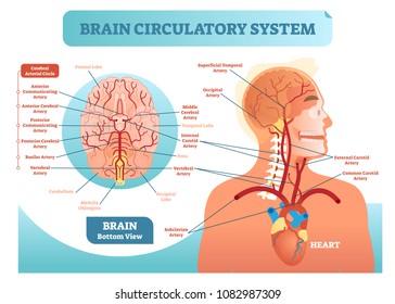
The arterial blood supply to the brain can be divided into the anterior and posterior circulation.
The blood vessels (and nerves) enter the brain through holes in the skull called foramina. There is a right sided aca and a left sided aca. Blood vessels labeled diagram, blood vessels labeling exercises, cat blood vessels labeled, human anatomy blood vessels, human blood. In this video i discuss the major arteries that supply the brain, starting with the internal carotid and vertebral arteries and covering many of the major. Sudden interruption of blood flow and oxygen to an area of brain tissue, which then may die (cerebrovascular accident, or cva, is another name for stroke.) ischemic stroke: An extraordinary degree of branching of blood vessels exists within the human body, which ensures that nearly every cell in the body lies within a short distance from at least one of. Blood supply to the brain is supplied by two main pairs of arteries, the internal carotid arteries and the vertebral arteries. Function and homeostasis of the brain relies on communication between its complex network of cells. The former is derived from the left and right internal carotid arteries, and the latter is haemorrhagic strokes occur when there is a rupture of a blood vessel or abnormal vascular structure within the brain. This vessel supplies blood to the front part of your brain, knows as your frontal lobe. • identification of blood vessels as arteries, capillaries or veins from the structure of their walls. Researchers have discovered how cells of the blood vessels sense the metabolic condition of the brain and alter vascular function in response. Veins return blood back toward the heart.
The dense tight junctions between endothelial cells prevent paracellular transport through the. Identify all of the blood vessels that are illustrated in the figure as you can while holding or otherwise examining whole brain specimens. In the cerebral medulla, the arteries and veins of the right side of the body are controlled from the left side of the brain; He says the restricted vessels prevent the blood from draining fast enough and injure the brain by causing a build up of iron which leads to ms. In the article on the ventricles within the cns, we will discuss their structure and.
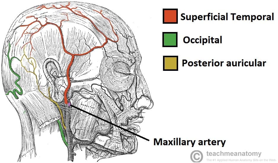
Major Arteries Of The Head And Neck Carotid Teachmeanatomy from teachmeanatomy.info Blood supply to the brain is supplied by two main pairs of arteries, the internal carotid arteries and the vertebral arteries. The only model that shows them is the somso torso. Ultrasound may offer a safe way to more as the name suggests, this is a barrier between the brain's blood vessels (capillaries) and the cells and other components that make up brain tissue. Blood flows throughout the body tissues in blood vessels, via bulk flow (i.e., all constituents together and in one direction). Examine a second specimen and notice any differences, such as asymmetries in the size of the vertebral or posterior communicating arteries. The difference in the structural characteristics of arteries, capillaries and veins is attributable to their respective functions. Scientists are developing new strategies for attaching drugs to molecules naturally transported across the barrier (labeled in green and blue). An extraordinary degree of branching of blood vessels exists within the human body, which ensures that nearly every cell in the body lies within a short distance from at least one of.
The 500 ms patients, both adults and children, also underwent mri scans of the brain to measure iron deposits in surrounding areas of the brain.
The difference in the structural characteristics of arteries, capillaries and veins is attributable to their respective functions. Blood vessels labeled diagram, blood vessels labeling exercises, cat blood vessels labeled, human anatomy blood vessels, human blood. The blood vessels are the components of the circulatory system that transport blood throughout the human body. Blood vessels are intricate networks of hollow tubes that transport blood throughout the entire body so that it can deliver valuable nutrients to and remove waste from cells. The 500 ms patients, both adults and children, also underwent mri scans of the brain to measure iron deposits in surrounding areas of the brain. If one of the major vessels becomes blocked, it is possible for blood flow to come across the circle of willis and prevent brain damage. Blood is also supplied to the brain by the vertebral a. Blood vessels in red in close communication with proliferating neuronal cells in the mouse cortex at embryonic day 10. Blood vessels flow blood throughout the body. The brain and its surrounding blood vessels exist in a close relationship. Supplies the posterior brain, blood supply to the entire brain is ensured by anastomoses between the vessels. There is a right sided aca and a left sided aca. Veins return blood back toward the heart.
Blood vessel endothelium is continuous with the inner tissue lining of organs such as the brain, lungs, skin, and heart. The two cell types ensure the integrity of the neural vasculature by maintaining the blood. Researchers have discovered how cells of the blood vessels sense the metabolic condition of the brain and alter vascular function in response. The former is derived from the left and right internal carotid arteries, and the latter is haemorrhagic strokes occur when there is a rupture of a blood vessel or abnormal vascular structure within the brain. The blood vessel wall is endowed with connective tissue, smooth muscle, and striated muscles.
Alila Medical Media Blood Supply Of The Brain Labeled Medical Illustration from d3e1m60ptf1oym.cloudfront.net Sudden interruption of blood flow and oxygen to an area of brain tissue, which then may die (cerebrovascular accident, or cva, is another name for stroke.) ischemic stroke: They also take waste and carbon dioxide away from the tissues. The former is derived from the left and right internal carotid arteries, and the latter is haemorrhagic strokes occur when there is a rupture of a blood vessel or abnormal vascular structure within the brain. In this video i discuss the major arteries that supply the brain, starting with the internal carotid and vertebral arteries and covering many of the major. The dense tight junctions between endothelial cells prevent paracellular transport through the. The carotid arteries and the vertebral arteries anterior cerebral artery (aca): This is particularly important structure due to its clinical implications, which are discussed in more detail in the article. Supplies the posterior brain, blood supply to the entire brain is ensured by anastomoses between the vessels.
The blood vessel wall is endowed with connective tissue, smooth muscle, and striated muscles.
The brain and its surrounding blood vessels exist in a close relationship. The two cell types ensure the integrity of the neural vasculature by maintaining the blood. Ultrasound may offer a safe way to more as the name suggests, this is a barrier between the brain's blood vessels (capillaries) and the cells and other components that make up brain tissue. Capillaries surround body cells and tissues to deliver and absorb oxygen, nutrients, and other substances. An extraordinary degree of branching of blood vessels exists within the human body, which ensures that nearly every cell in the body lies within a short distance from at least one of. This vessel supplies blood to the front part of your brain, knows as your frontal lobe. These vessels transport blood cells, nutrients, and oxygen to the tissues of the body. Internal carotid artery (anterior circulation), vertebral artery (posterior circulation), and their hexagonal anastomotic network called blood brain barrier refers to the wall between the brain tissue and blood vessels. The dense tight junctions between endothelial cells prevent paracellular transport through the. Label the blood vessels of the male pelvis using the hints provided. The blood vessels (and nerves) enter the brain through holes in the skull called foramina. In this video i discuss the major arteries that supply the brain, starting with the internal carotid and vertebral arteries and covering many of the major. In the cerebral medulla, the arteries and veins of the right side of the body are controlled from the left side of the brain;
Our blood vessel models have the same elasticity as the living body, greater tensile strength and a blood vessels labeled. Blood vessels flow blood throughout the body.


0 Comments