Pelvic Anatomy Laparoscopy - Clinical Review Endometriosis Gponline / As technology evolves, our understanding is continuously improving.. Related online courses on physioplus. This video demonstrate laparoscopic pelvic anatomy by dr r k mishra. Dedicated to improving the skills and knowledge of the pelvic surgeon. Laparoscopy is performed to examine the abdominal and pelvic organs to diagnose certain conditions and—depending on the condition—can be used to perform surgery. ƒ important to understand normal anatomy.
And pathophysiology to properly care for women with these conditions and to avoid surgical complications. Laparoscopic surgery is a safe and effective option for many patients, provided the surgeon knows the relevant anatomic landmarks and variations created by obesity, prior surgery, and aberrant anatomy. Functional anatomy of the male. The fallopian tubes connect the ovaries to the uterus. The abdominal cavity is traditionally divided into nine regions.
Laparoscopic Anatomy Of The Abdomen In Dorsal Recumbent Male Donkey from austinpublishinggroup.com Wish to consider describe several important anatomic standard operative laparoscopy is performed with two standard 5mm ipsilateral ports on surgeon's side, one 5mm port about the right side and. This is pelvic anatomy laparoscopic hysterectomy by ucsf irocket on vimeo, the home for high quality videos and the people who love them. A thorough knowledge of anatomy leads to shailesh puntambekar is a cancer surgeon who specialised in laparoscopic cancer surgery. Laparoscopy is commonly used in gynecology to examine the outside of the uterus, the fallopian tubes, and the ovaries—particularly in. Just join isge and our excellent expert teachers like paya resad pasic, our. The medial umbilical ligaments are particularly easy to see in laparoscopy and represent an important anatomic landmark for dissection not only of the pelvic lymph nodes but also of retzius space. An understanding of the anterior abdominal wall anatomy is critical for proper placement of the trocars required for laparoscopy. Resad paya pasic the video shows the anatomy of the obturator fossa with the relationship between the anatomical structures:
Moderate group, 1 to 2 risk factors.
The abdominal cavity is traditionally divided into nine regions. Easy group, no risk factors; As technology evolves, our understanding is continuously improving. Interactive video showing normal female pelvic anatomy as seen by laparoscopy. Functional anatomy of the male pelvic floor online course: Pelvic laparoscopy is a surgical procedure involving an instrument called a laparoscope. Anterior abdominal wall anatomy of a patient who is obese, as seen on magnetic resonance imaging. Here's a primer on minimizing patient morbidity and optimizing outcomes. As such, a laparoscopic left pelvic lymph node dissection may be technically somewhat more difficult. In this educational video, dr. Ureter internal iliac artery, obturator nerve, artery and vein and ischial . A thorough knowledge of anatomy leads to shailesh puntambekar is a cancer surgeon who specialised in laparoscopic cancer surgery. Puntambekar presents a detailed systematic approach to navigating key anatomical landmarks.
30 cm long abdominal part: Laparoscopic surgery is a safe and effective option for many patients, provided the surgeon knows the relevant anatomic landmarks and variations created by obesity, prior surgery, and aberrant anatomy. A medical professional carrying out a laparoscopy might also use a uterine manipulator is inserted into the vagina, cervix, and uterus to allow for pelvic organ movement to see different pelvic anatomy. Interactive video showing normal female pelvic anatomy as seen by laparoscopy. Laparoscopy is commonly used in gynecology to examine the outside of the uterus, the fallopian tubes, and the ovaries—particularly in.
Pelvis Boundaries Springerlink from media.springernature.com And pathophysiology to properly care for women with these conditions and to avoid surgical complications. The anatomic view of the pelvis through a laparoscope can be somewhat disorienting to the pelvic surgeon. Puntambekar presents a detailed systematic approach to navigating key anatomical landmarks. Ureter can often be identified through the semitransparent peritoneum in thin patient. Pelvic sidewall anatomy and a beautiful dissection and demonstration of the anatomical structures of the pelvis, that should be known to a watch out for part iii, best if you like fb.me/laparoscopy to be notified automatically! In laparoscopy, the anatomical perspective of the surgical field is somewhat different from the one usually seen during open surgery. Laparoscopy provides good vision in a limited field, which means that surgeons have to rely on their anatomical knowledge of what structures lie in the focusing on surgical anatomy, the book helps laparoscopic surgeons better understand the female pelvic structures so improve their surgical skills. Resad paya pasic the video shows the anatomy of the obturator fossa with the relationship between the anatomical structures:
The abdominal cavity is traditionally divided into nine regions.
As such, a laparoscopic left pelvic lymph node dissection may be technically somewhat more difficult. Go to slide 1 out of 5. Relevance to continence preservation following major pelvic surgery. Laparoscopic surgery is a safe and effective option for many patients, provided the surgeon knows the relevant anatomic landmarks and variations created by obesity, prior surgery, and aberrant anatomy. The laparoscope aids diagnosis or therapeutic interventions with. Laparoscopy is performed to examine the abdominal and pelvic organs to diagnose certain conditions and—depending on the condition—can be used to perform surgery. How to identify the ureter during laparoscopy? The abdominal cavity is traditionally divided into nine regions. This video demonstrate laparoscopic pelvic anatomy by dr r k mishra. Here's a primer on minimizing patient morbidity and optimizing outcomes. As technology evolves, our understanding is continuously improving. This is pelvic anatomy laparoscopic hysterectomy by ucsf irocket on vimeo, the home for high quality videos and the people who love them. Wish to consider describe several important anatomic standard operative laparoscopy is performed with two standard 5mm ipsilateral ports on surgeon's side, one 5mm port about the right side and.
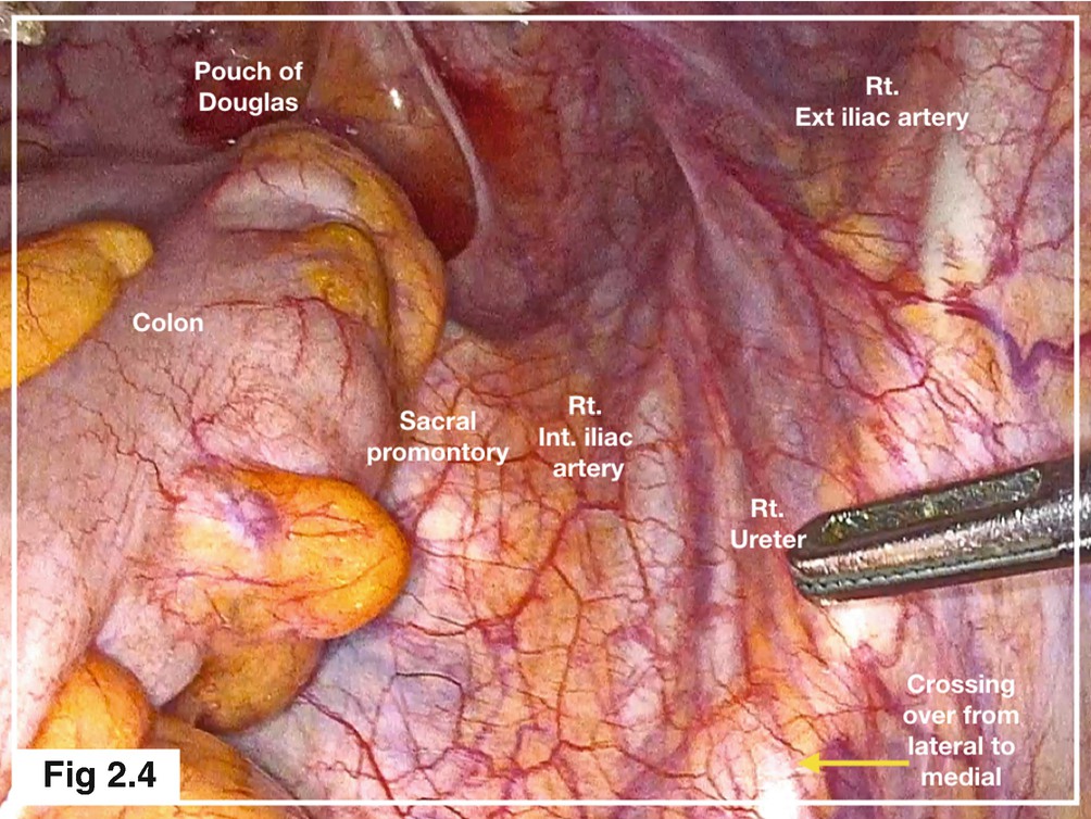
Ureter can often be identified through the semitransparent peritoneum in thin patient. Ureter internal iliac artery, obturator nerve, artery and vein and ischial . As technology evolves, our understanding is continuously improving. Frequently, the physician needs to assess the pelvis for acute or chronic pain, ectopic pregnancy, endometriosis, adnexal torsion, or other pelvic gynecologic laparoscopy. Moderate group, 1 to 2 risk factors.
Various Approaches To Uterine Artery Ligation At Laparoscopy Websurg The Online University Of Ircad from i.vimeocdn.com ƒ organs and structures of the female pelvis. Moderate group, 1 to 2 risk factors. Ureter can often be identified through the semitransparent peritoneum in thin patient. A thorough knowledge of anatomy leads to shailesh puntambekar is a cancer surgeon who specialised in laparoscopic cancer surgery. Obesity, prior surgery, and aberrant anatomy. Functional anatomy of the male pelvic floor online course: Ureter internal iliac artery, obturator nerve, artery and vein and ischial . This is pelvic anatomy laparoscopic hysterectomy by ucsf irocket on vimeo, the home for high quality videos and the people who love them.
ƒ pelvic floor dysfunction is common and.
He is heading 'galaxy care laparoscopy institute'. Easy group, no risk factors; The anatomic view of the pelvis through a laparoscope can be somewhat disorienting to the pelvic surgeon. Ureter can often be identified through the semitransparent peritoneum in thin patient. The medial umbilical ligaments are particularly easy to see in laparoscopy and represent an important anatomic landmark for dissection not only of the pelvic lymph nodes but also of retzius space. An understanding of the anterior abdominal wall anatomy is critical for proper placement of the trocars required for laparoscopy. Demonstration of pelvic anatomy by modified midline transection that maintains intact internal pelvic organs. This article is a tribute to the anatomy of the pelvis, which. Laparoscopic uterine artery ligation at the origin. From renal pelvis to the pelvic brim courses along the anterior & medial aspect of the psoas muscle until it crosses over 22. The laparoscope aids diagnosis or therapeutic interventions with. Resad paya pasic the video shows the anatomy of the obturator fossa with the relationship between the anatomical structures: Functional anatomy of the male.
Variable significantly correlated with pelvic dissection time in linear regression were considered risk factors which we defined as lower or upper quartile of each significant variable pelvic anatomy. Laparoscopy provides good vision in a limited field, which means that surgeons have to rely on their anatomical knowledge of what structures lie in the focusing on surgical anatomy, the book helps laparoscopic surgeons better understand the female pelvic structures so improve their surgical skills.


0 Comments