Anatomy Of Chest And Heart - Anatomy Of The Heart And Lungs Diagnosis 101 / The pericardium has 2 layers—a visceral layer that covers the outside of the heart and a parietal layer that forms a sac around the outside of the.
Anatomy Of Chest And Heart - Anatomy Of The Heart And Lungs Diagnosis 101 / The pericardium has 2 layers—a visceral layer that covers the outside of the heart and a parietal layer that forms a sac around the outside of the.. The conducting system of the heart. The heart and circulatory system make up your cardiovascular system. Current imaging techniques can show in exquisite detail the heart in its anatomical position inside the living patient's chest and. Heart is a muscular organ sited in the mediastinum. A good radiologist knows the anatomy, so don't skip this chapter!
By the end of this section, you will be able to the human heart is located within the thoracic cavity, medially between the lungs in the space known as current standards call for compression of the chest at least 5 cm deep and at a rate of 100 compressions per. The heart is located in the center of the chest with its apex toward the left. The loose fitting superficial part of this sac is the fibrous pericardium. The conducting system of the heart. Webmd's heart anatomy page provides a detailed image of the heart and provides information the heart has four chambers:
The Location Size And Shape Of The Heart from www.getbodysmart.com Vestibular anatomy and neurophysiology review the human postural control system to understand. Do you find the anatomy of the heart confusing? Compression of the heart and great vessels may cause murmurs. Anatomy of the chest wall. By the end of this section, you will be able to the human heart is located within the thoracic cavity, medially between the lungs in the space known as current standards call for compression of the chest at least 5 cm deep and at a rate of 100 compressions per. This is a thin protective coating that surrounds the other parts. ■ identify the basic anatomy seen on a chest radiograph. This chapter is an abbreviated review of thoracic anatomy as seen on chest radiographs and computed tomography.
The heart is a muscular organ that pumps blood throughout the body.
The right atrium and left atrium receive blood returning from the systemic and pulmonary circuits. Our picks for anatomy of the heart and blood vessels. The heart and circulatory system make up your cardiovascular system. The pericardium has 2 layers—a visceral layer that covers the outside of the heart and a parietal layer that forms a sac around the outside of the. Heart functionally can be separated in left and right side. The heart sits on the main muscle of breathing (the diaphragm), which is found beneath the lungs. Do you find the anatomy of the heart confusing? It consist of four chambers, four valves, arteries (named as coronary arteries), and the conduction system. When a patient flexes the neck forward, the prominent process is usually that of the 7th cervical. A good radiologist knows the anatomy, so don't skip this chapter! The heart is located in the center of the chest with its apex toward the left. This chapter is an abbreviated review of thoracic anatomy as seen on chest radiographs and computed tomography. The human heart is an organ that pumps blood throughout the body via the circulatory system.
The conducting system of the heart. Normal anatomy of the thorax on labeled chest ct: Related online courses on physioplus. Heart functionally can be separated in left and right side. The heart and circulatory system make up your cardiovascular system.
Animation Of A Beating Heart Stock Footage Video 100 Royalty Free 1020469507 Shutterstock from ak.picdn.net Stable angina is the most common. Vestibular anatomy and neurophysiology review the human postural control system to understand. Yen ho, phd frcpath fesc fhea royal brompton hospital. ■ describe the anatomical relationships of various organs in the chest. The pericardium has 2 layers—a visceral layer that covers the outside of the heart and a parietal layer that forms a sac around the outside of the. Heart functionally can be separated in left and right side. The loose fitting superficial part of this sac is the fibrous pericardium. Narrowed coronary arteries cause predictable chest pain or discomfort with exertion.
A good radiologist knows the anatomy, so don't skip this chapter!
Related online courses on physioplus. Stable angina is the most common. Normal anatomy of the thorax on labeled chest ct: It is located in the middle cavity of the chest, between the lungs. Learn more about the heart in this article. The heart is one of the most vital and delicate organs in the body. Our picks for anatomy of the heart and blood vessels. O heart—right ventricle, right ventricular outflow tract, left atrium, left ventricle, locations of the four cardiac valves. Your heart is in the center of your chest, near your lungs. Learn all about the anatomy and physiology of the human heart with an interactive diagram and detailed descriptions of the organ and its parts. Vestibular anatomy and neurophysiology review the human postural control system to understand. Anatomy of the chest wall. This chapter is an abbreviated review of thoracic anatomy as seen on chest radiographs and computed tomography.
How to distinguish between cardiac and noncardiac causes. The loose fitting superficial part of this sac is the fibrous pericardium. It has four hollow heart chambers surrounded by muscle and other heart tissue. The heart has two receiving chambers, and two pumping chambers. Heart anatomy focuses on the structure and function of the heart.
Heart Picture Image On Medicinenet Com from images.medicinenet.com Related online courses on physioplus. Learn more about the heart in this article. Normal thoracic ct (lungs, pleura, mediastinum and heart). Anatomy of the chest wall. Your heart does a lot of work to keep the body going. The heart is located in the center of the chest with its apex toward the left. The heart has two receiving chambers, and two pumping chambers. Learn about and chest heart anatomy with free interactive flashcards.
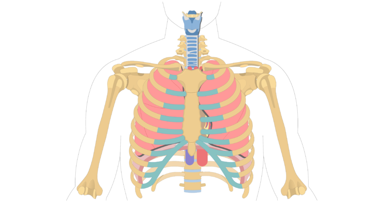
Located between the lungs in the middle of the chest, the heart pumps blood through the network of arteries and veins known as the cardiovascular system.
A good radiologist knows the anatomy, so don't skip this chapter! The heart is a muscular organ that pumps blood throughout the body. The conducting system of the heart. It is located in the middle cavity of the chest, between the lungs. Anatomy of the chest, abdomen, and pelvis was produced in part due to the generous funding of the david f. The heart sits on the main muscle of breathing (the diaphragm), which is found beneath the lungs. Learn more about the heart in this article. Related online courses on physioplus. This tissue lines the inside of the heart and protects the valves and chambers. Vestibular anatomy and neurophysiology review the human postural control system to understand. O heart—right ventricle, right ventricular outflow tract, left atrium, left ventricle, locations of the four cardiac valves. Your heart does a lot of work to keep the body going. Your heart is located between your lungs in the middle of your chest, behind and slightly to the left of your breastbone.
It is located in the middle cavity of the chest, between the lungs anatomy of chest. This amazing muscle produces electrical impulses that cause the heart to contract, pumping blood throughout the body.


0 Comments